Implant Failure Leading to Union in a Case of Delayed union of Distal Femur Fracture – Implant Failures are not always Failures
Vol 1 | Issue 1 | Jan-Apr 2016 | page: 17-19 | Vikram V. Kadu[1], K. A. Saindane[1],Ninad Goghate[1], Neha N Godghate[1].
Author: Vikram V. Kadu[1], K. A. Saindane[1],Ninad Goghate[1], Neha N Godghate[1].
[1] ACPM Medical College, Dhule – 424001, Maharashtra, India
Address of Correspondence
Dr. Vikram V. Kadu
ACPM Medical College, Dhule – 424001, Maharashtra India
Email : vikram1065@gmail.com
Abstract
Introduction: Supracondylar fractures of the femur are seen in young adults, usually as a result of high-energy trauma and in elderly individuals with osteoporotic bones. These fractures may have extension to intercondylar region or may be comminuted. In order to achieve early range of motion these fractures need anatomical and rigid fixation. 95 Dynamic condylar screw being a strong implant with rigid construct provides good rigid and stable fixation. Implant failure generally results from improper reduction, inadequate fixation or early mobilization after the surgery.
Case report: 42 yrs old male suffered RTA and was brought to our OPD with complaints of pain over left knee and distal femur region. He had supracondylar femur with intercondylar extension. He was kept on skeletal traction and then operated with 95 DCS. Post-operative radiographs were satisfactory showing good reduction and adequate fixation. There was slight distraction at the fracture site which was acceptable. At 5 months follow-up, radiographs showed signs of delayed union. At 8 months follow-up, radiograph showed implant failure and collapse at the fracture site. Implant was kept in situ and weight bearing was continued. Fracture united completely with functional range of motion.
Conclusion: Broken implant is not a sign of broken biology. Careful assessment of fracture should be done before deciding for revision surgery
Keywords: Distal femur fracture, implant failure, bony union.
Introduction
Jthe increasing lifetime of the population on a world-wide scale over the last decades has led to a significant growth in the use of surgical implants in affected patients. Orthopedic implants can be defined as devices that are used to replace bones or to help the fixation of fractured bones. Since the average human life expectation has increased significantly in the past decades and is supposed to keep increasing in the near future [1,2], more and more implants are being used. Supracondylar fractures of the femur are often difficult to treat. These fractures require careful management to obtain good cosmetic and functional results. The main problem is obtaining and maintaining adequate reduction of both shaft and articular fragments while allowing function of the knee to be regained at an early stage. Dynamic Condylar Screw is easier to insert, provide more inter- fragmentary compression across an intercondylar fracture and surgical treatment of supracondylar fractures.[3,4] However, an internal fixation device may fail to hold a reduced fracture until union, giving rise to non-union or delayed union. Implant failures arise mainly from loosening or breakage of the internal fixation device. Because bones are more flexible than metal plates, screwing a metallic plate to bone stiffens it and produces ”stress riser” at each end of the plate. In the absence of union, even the strongest metal plates and screws will eventually break or pull out of bone[5].
Case Presentation
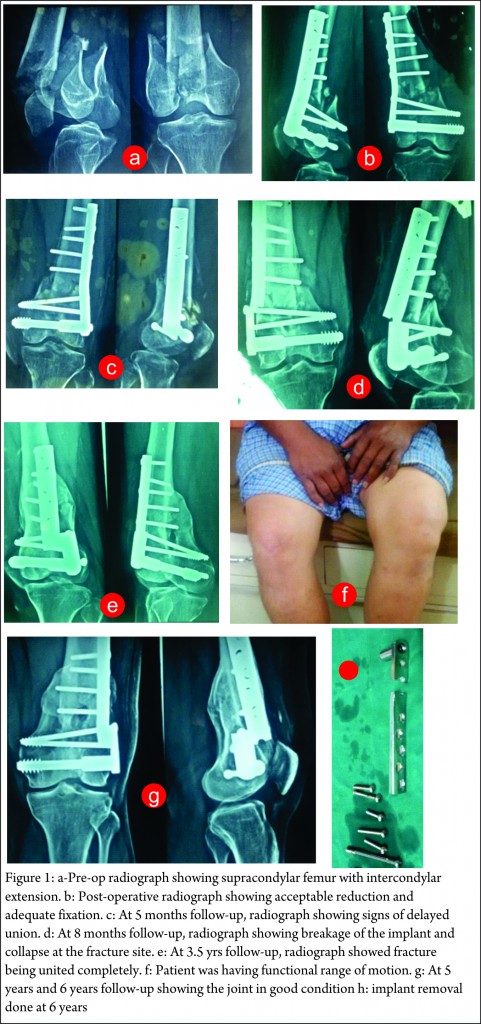
42 yrs old male suffered RTA and was brought to our OPD with complaints of pain over left knee and distal thigh region. Radiograph showed supracondylar femur with intercondylar extension (Fig 1a). He was given proximal tibial skeletal traction to achieve length and maintain reduction till he was medically fit for surgery. Definitive management consisted of fixation of the fracture fragments with 95 degree DCS. Post-operative radiographs were satisfactory showing good reduction and adequate fixation (Fig 1b). At 2.5 months follow-up, radiographs showed slight distraction at the fracture site which was acceptable. Partial weight bearing was started. At 5 months follow-up, radiographs showed signs of delayed union (Fig 1c). Full weight bearing was started. At 8 months follow-up, radiograph showed breakage of the implant and collapse at the fracture site (Fig 1d). Implant was kept in situ and weight bearing was continued. Further collapse at the fracture site due to weight bearing resulted in union of fracture. At 3.5 yrs follow-up, radiographs showed fracture being united completely (Fig 1e) and patient had functional range of motion (Fig 1f). The patient was followed up every 6 months and was assessed radiologically and clinically. At 6 yrs follow up the bone was well healed and implant was removed (Fig 1g-h). Presently patient is walking without any support and has functional range of motion.
Discussion
Fractures in the distal femur have posed considerable therapeutic challenges [6,7] throughout the history of fracture treatment.[8,9,10] Supracondylar distal femoral fractures may be classified as extraarticular, unicondylar, or bicondylar, and the fractures may have an intercondylar extension.[11,1 2] The supracondylar region of the femur represents the metaphyseal transition between the femoral diaphysis and the distal femoral articular surface. Within this region the geometry and the function of the bone changes from weight bearing to articulation. The alignment of the femoral shaft becomes an important consideration when dealing with the supracondylar femur fractures. Many poor results stem from a failure to realign the mechanical axis (which passes through the head of the femur and the middle of the knee joint) and poor reconstruction of the anatomic axis which has an average valgus angulation of 9 degrees relative to the vertical axis. The aim of fracture treatment is to achieve union and timely functional recovery. Plates and screws are known to rigidly fix fractures which in turns slow down the rate of callus formation. The success of an implant depends on multiple factors and is necessary to determine whether failure was inherent to the device or was caused by external factors such as installation, patient co-operation or rate of fracture healing.[13] “Dynamic Condylar Screw” is technically easier to apply than a blade plate. It allows adjustment in the sagittal plane and moreover it can be used for both supracondylar and intercondylar fracture with at least 4 cm intact bone in the femoral condyles above the intercondylar notch is necessary for successful fixation .[14,15,16] Each lower limb bears an average of three times the body weight at the stance phase of the gait cycle. It is expected that early weight bearing before significant union may lead to loosening or fatigue failure of implants. The supracondylar fracture femur in our case was fixed adequately but showed delayed union and could have gone into non-union requiring another surgery consisting of secondary bone grafting. Patient was allowed weight bearing in order to achieve compression at the fracture site. During the weight bearing period the implant broke and fracture fragments collapsed. The collapse of the fragments resulted in the compression at the fracture site further leading to formation of exuberant callus and solid union of the fracture. This would not have been possible without implant failure. Implant failure though considered as one of the complications in orthopaedic surgeries and requires revision of surgery with another implant, sometime it can sometimes give surprising outcome. However there is no method to predict such outcome and probably our patient was simply lucky.
Conclusion
95 DCS is a good modality for treating supracondylar fracture femur. Some fractures may go into delayed union. Implant failure in cases of delayed union results in collapse at the fracture site leading to approximation of the fracture fragments and may be fracture union. The message from this report is that broken implant is not really a sign of broken biology. We should carefully assess the local mileu and decide on revision surgery
Clinical Message
Implant failure though considered as one of the complications in orthopaedic surgeries, sometimes it can have surprising outcome. Probably our patient was lucky, but we also believe that broken implant is not a definative indication of broken biology. One should never hurry in revising the surgery, instead assessment of local factors should be done to take decision on revision surgery
References
1. Ali MA, Shafique M, Shoaib M. Fixation of femoral supracondylar fractures by dynamic condylar screw. Med Channel 2004; 10:65-7.
2. Fini, M., Aldini, N.N., Torricel1i, P., Giavaresi, G., Borsari, V., Lenger, H., Bernauer, J., Giardino, R., Chiesa, R, Cigada, A: A new austenitic stainless steel with negligible nickel content: an in vitro and in vivo comparative investigation. Biomaterials 24, 4929–4939 (2003).
3. Jeon IH, Oh CW, Kim SJ, Park BC, Kyung HS, Ihn JC. Minimally invasive percutaneous
plating of distal femoral fractures using the dynamic condylar screw. J Trauma 2004; 57:1 048-52.
4.Lumley JSP, Craver JC. Complications of fracture healing. Surg Int 200; 54:206.
5.David J. Dandy, Dennis J. Edwards. Essential Orthopaedics and Trauma. 4th edition. Churchill Livingstone: Edinburgh. 2003; 45 & 77
6.Christodoulou A, Terzidis I, Ploumis A, Metsovitis S, Koukoulidis A, Toptsis C. Supracondylar femoral fractures in elderly patients treated with the dynamic condylar screw and the retrograde intramedullary nail: a comparative study of the two methods. Arch Orthop Trauma Surg 2005; 125:73-9.
7.Seligson D, Mulier T, Keirsbilck S, Been J. supracondylar femur fractures. J Orthop Plating of femoral shaft fractures: a review of 15 cases. Acta Orthop Belg 2001; 67:24-31 .
8.Neer CS, Grantham S A, Shelton ML. Supracondylar fracture of the adult femur. J Bone Joint Surg 1967; 49:591 -61 3.
9.Schatzker J, Lambert DC. Supracondylar fractures of the femur. Clin Orthop fixation of distal femoral fractures: results 1979; 138:77-83.
10.Stewart MJ, Sisk TD, Wallace SL. Fractures of the distal third of the femur. J Bone Joint Surg 1966; 48:784-807
11.Rogers LF. The hip and emoral shaft. In: Rogers LF, ed. Radiology of skeletal trauma, 2nd ed. New York: Churchill Livingstone, 1992:1199–1317
12.Helfet DL. Fractures of the distal femur. In: Browner BD, Jupiter JB, Levine AM, Trafton PG, eds. Skeletal trauma. Philadelphia: Saunders, 1992:1643–1683
13.Christain CA. Campbell Operative Orthopaedics. 9th Edition. Mosby Publishers, 1998; 2000-01
14.Gregory P, Sanders R. The treatment of supracondylar-intracondylar fractures of femur using the dynamic condylar screw. Tech Orthop 1995; 9:1 95.
15.Harder Y, Martinet O, Barraud GE. The mechanics of internal fixation of fractures of the distal femur: a comparison of the condylar screw (CS) with the condylar plate (CP). Injury 1999; 30:31.
16.Schatzker J, Mahomed N, S chiffman K, Kellam J. Dynamic condylar screw. J Bone Joint Surg Br 1989; 74: 1 22-5.
| How to Cite this article: Kadu VV, Saindane KA, Goghate N, Godghate NN. Implant Failure Leading to Union in a Case of Delayed union of Distal Femur Fracture – Implant Failures are not always Failures. Journal of Orthopaedic Complications Jan-April 2016; 1(1):17-19. |

