Narrative Review: Arthroscopic-Assisted Percutaneous Fixation of Calcaneus Fractures
Review Article | Vol 2 | Issue 3 | September-December 2025 | page: 08-11 | Sachin Kale, Sandeep Deore, Kaustav Das, Atul Yadav, Daniyal Shaikh, Nikhil Makhija, Shivesh Datta
Submitted Date: 08-05-2025, Review Date: 11-06-2025, Accepted Date: 21-08-2025 & Published Date: 10-12-2025
https://doi.org/10.13107/joc.2025.v02.i03.34
Authors: Sachin Kale [1], Sandeep Deore [1], Kaustav Das [1], Atul Yadav [1], Daniyal Shaikh [1], Nikhil Makhija [1], Shivesh Datta [1]
[1] Department of Orthopaedics, D.Y Patil School of Medicine and Hospital, Navi Mumbai, Maharashtra, India.
Address of Correspondence
Dr. Sachin Kale,
Department of Orthopaedics, D.Y Patil School of Medicine and Hospital, Navi Mumbai, Maharashtra, India.
E-mail: sachinkale@gmail.com
Abstract
Calcaneus fractures are the most prevalent tarsal bone injuries, often resulting from high-energy trauma. These injuries pose significant challenges in orthopaedic management due to the complex anatomy of the calcaneus and the potential for severe soft tissue complications. Traditional open reduction and internal fixation (ORIF) methods, while effective, are associated with notable risks, including wound complications and infections. Recently, arthroscopic-assisted percutaneous fixation has emerged as a minimally invasive alternative, offering the potential for reduced complications and improved outcomes. This narrative review explores the technique’s methodology, advantages, limitations, and its evolving role in the treatment of calcaneus fractures.
Keywords: Calcaneus fractures, Arthroscopy, Percutaneous fixation, Minimally invasive surgery, Orthopaedic trauma
Introduction
Calcaneus fractures account for approximately 60% of tarsal bone injuries and are predominantly caused by high-energy mechanisms such as falls from height or motor vehicle accidents [1, 2]. The calcaneus’s intricate structure and articulation with the talus and cuboid bones necessitate precise anatomical reduction and fixation to restore function and prevent long-term complications like subtalar arthritis [3]. Traditional ORIF has been the gold standard for displaced intra-articular fractures; however, it carries a significant risk of soft tissue complications due to the extensive surgical exposure required [4].
Arthroscopic-assisted percutaneous fixation has emerged as a promising alternative, offering enhanced visualization and reduced soft tissue disruption [5]. This technique utilizes the advantages of arthroscopy to inspect and address intra-articular pathology while employing minimally invasive percutaneous methods for fracture stabilization.
Methodology
Preoperative Planning
Effective management of calcaneus fractures begins with comprehensive preoperative planning. Advanced imaging techniques, particularly CT scans, are critical for understanding fracture patterns, degree of displacement, and articular involvement [6]. This imaging helps in assessing the extent of displacement and articular involvement, which are crucial for surgical decision-making.


Surgical Technique
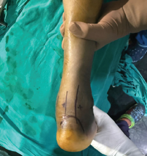
The arthroscopic-assisted percutaneous fixation technique involves a combination of endoscopic visualization and minimally invasive hardware placement. Patients are positioned supine, with the affected limb elevated to facilitate access to the lateral side of the foot. Regional or general anesthesia is typically used [7].

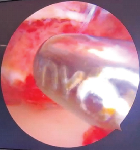
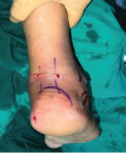
• Arthroscopic Assessment: Standard anterolateral and posterolateral portals are established to introduce the arthroscope into the subtalar joint. This allows for direct visualization of the joint surfaces and fracture lines, enabling precise assessment of intra-articular pathology [8].
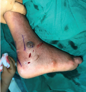
• Fracture Reduction: Under arthroscopic guidance, reduction is achieved using percutaneous tools such as Schanz pins or Kirschner wires to manipulate fragments into alignment while confirming joint surface congruity through the arthroscope [9].
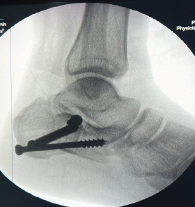
• Percutaneous Fixation: Once reduction is confirmed, percutaneous cannulated screws or Kirschner wires are inserted to stabilize the fracture. Fluoroscopy is used to verify correct placement and alignment of the hardware [10].
• Postoperative Care: Patients are typically immobilized in a boot or cast with non-weight-bearing instructions, progressing to weight-bearing as tolerated. Rehabilitation focuses on restoring range of motion and strength [11].
Discussion
The use of arthroscopy in fracture management provides significant advantages over traditional methods. By minimizing soft tissue disruption, this approach reduces the risk of complications such as wound dehiscence and infection [12]. Arthroscopic visualization allows for precise reduction of the articular surface, which is critical for optimal outcomes [13]. The minimally invasive nature of the procedure also contributes to faster recovery times and earlier mobilization [14].
However, the technique is not without its challenges. It requires a high level of skill in both arthroscopy and fracture fixation, presenting a steep learning curve for surgeons [15]. Additionally, the presence of significant swelling or hematoma can complicate visualization, necessitating careful intraoperative management [16].
Potential complications specific to this technique include the risk of neurovascular injury during portal placement, hardware-related issues such as screw back-out or migration, and potential for inadequate reduction due to limited visibility in complex fracture patterns [17].
Results
Clinical outcomes following arthroscopic-assisted percutaneous fixation have been promising. Studies report high union rates, with most patients returning to pre-injury activity levels within three months [18]. Complications are generally minimal, with significantly fewer wound-related issues compared to traditional ORIF [19]. Long-term follow-up indicates satisfactory functional outcomes with low rates of post-traumatic arthritis [20].
Conclusion
Arthroscopic-assisted percutaneous fixation represents a significant advancement in the management of calcaneus fractures. By combining the benefits of minimally invasive surgery with enhanced visualization, this approach offers the potential for improved patient outcomes and reduced complications. Ongoing research and experience will continue to refine this technique and establish its role in orthopaedic trauma surgery.
Learning Points
• Arthroscopic techniques provide superior visualization for fracture reduction.
• Minimally invasive approaches decrease soft tissue complications and enhance recovery.
• Surgeons must be proficient in both arthroscopy and percutaneous fixation techniques to achieve the best outcomes.
References
1. Sanders R. Displaced intra-articular fractures of the calcaneus. J Bone Joint Surg Am. 2000;82(2):225-250.
2. Buckley R, Tough S, McCormack R, et al. Operative compared with nonoperative treatment of displaced intra-articular calcaneal fractures. J Bone Joint Surg Am. 2002;84(10):1733-1744.
3. Thordarson DB, Krieger LE. Operative vs. nonoperative treatment of intra-articular fractures of the calcaneus: a prospective randomized trial. Foot Ankle Int. 1996;17(1):2-9.
4. Gavlik JM, Rammelt S, Zwipp H. Percutaneous arthroscopic treatment of intra-articular calcaneus fractures. Arch Orthop Trauma Surg. 2002;122(8):424-428.
5. Rammelt S, Zwipp H. Fractures of the calcaneus: current treatment strategies. Acta Chir Orthop Traumatol Cech. 2014;81(3):177-196.
6. Schepers T, van Lieshout EM, Heetveld MJ, et al. Current concepts in the treatment of intra-articular calcaneal fractures: results of a nationwide survey. Int Orthop. 2008;32(5):711-715.
7. Schepers T. The use of a locking, minimally invasive, percutaneous plate osteosynthesis technique for the treatment of displaced intra-articular calcaneal fractures. J Orthop Trauma. 2011;25(7):e75-e80.
8. Zhang Y, Li S, Wu H, et al. Minimally invasive treatment of intra-articular calcaneal fracture by percutaneous leverage, anatomical plate, and compression bolts: a cohort study. Int J Surg. 2014;12(7):645-650.
9. Ebraheim NA, Elgafy H, Sabry FF, et al. Sinus tarsi approach with trans-articular fixation for displaced intra-articular fractures of the calcaneus. Foot Ankle Int. 2000;21(2):105-113.
10. Benirschke SK, Sangeorzan BJ. Extensive intraarticular fractures of the foot. Surgical management of calcaneal fractures. Clin Orthop Relat Res. 1993;(292):128-134.
11. Folk JW, Starr AJ, Early JS. Early wound complications of operative treatment of calcaneus fractures: analysis of 190 fractures. J Orthop Trauma. 1999;13(5):369-372.
12. Rammelt S, Amlang M, Barthel S, et al. Minimally-invasive treatment of calcaneal fractures. Injury. 2004;35(2):55-63.
13. Hohendorff B, Sander K, Muhr G, et al. Long-term results after operative treatment for intra-articular fractures of the calcaneus. J Orthop Trauma. 2007;21(10):643-648.
14. Xia S, Liu J, Yan L, et al. Treatment of calcaneal fractures through a minimally invasive sinus tarsi approach combined with medial distraction technique. BMC Musculoskelet Disord. 2017;18(1):108.
15. Shi F, Wu X, Liu H, et al. Minimally invasive sinus tarsi approach with percutaneous plate and screw fixation for intra-articular calcaneal fractures. J Orthop Surg Res. 2016;11(1):164.
16. Gavlik JM, Rammelt S, Zwipp H. Percutaneous arthroscopic treatment of intra-articular calcaneus fractures. Arch Orthop Trauma Surg. 2002;122(8):424-428.
17. Rammelt S, Zwipp H. Fractures of the calcaneus: current treatment strategies. Acta Chir Orthop Traumatol Cech. 2014;81(3):177-196.
18. Gavlik JM, Rammelt S, Zwipp H. Percutaneous arthroscopic treatment of intra-articular calcaneus fractures. Arch Orthop Trauma Surg. 2002;122(8):424-428.
19. Rammelt S, Zwipp H. Fractures of the calcaneus: current treatment strategies. Acta Chir Orthop Traumatol Cech. 2014;81(3):177-196.
20. Benirschke SK, Sangeorzan BJ. Extensive intraarticular fractures of the foot. Surgical management of calcaneal fractures. Clin Orthop Relat Res. 1993;(292):128-134.
| How to Cite this article: Kale S, Deore S, Das K, Yadav A, Shaikh D, Makhija N, Datta S. Narrative Review: Arthroscopic-Assisted Percutaneous Fixation of Calcaneus Fractures. Journal of Orthopaedic Complications. September-December 2025;2(3):08-11. |